m6A-centered Crosstalk Information
Mechanism of Crosstalk between m6A Modification and Epigenetic Regulation
| Crosstalk ID |
M6ACROT05346
|
[1], [2] | |||
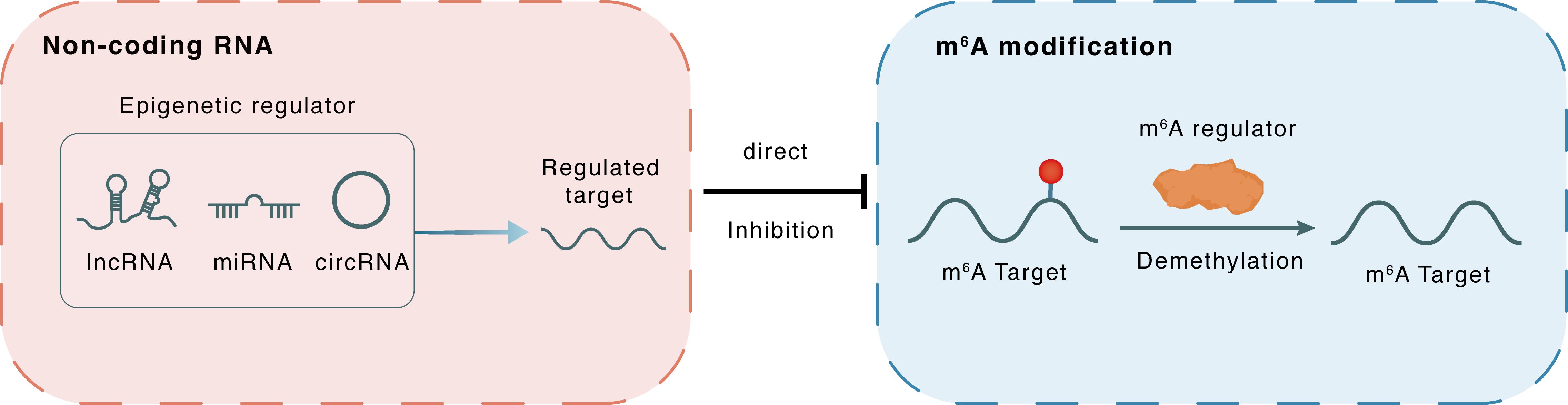 Non-coding RNA
MiR-155
FTO
lncRNA miRNA circRNA
Direct
Inhibition
m6A modification
PPARGC1A
PPARGC1A
FTO
Demethylation
Non-coding RNA
MiR-155
FTO
lncRNA miRNA circRNA
Direct
Inhibition
m6A modification
PPARGC1A
PPARGC1A
FTO
Demethylation
 : m6A sites : m6A sites
|
|||||
| m6A Modification: | |||||
|---|---|---|---|---|---|
| m6A Regulator | Fat mass and obesity-associated protein (FTO) | ERASER | |||
| m6A Target | PPAR-gamma coactivator 1-alpha (PGC-1a/PPARGC1A) | ||||
| Epigenetic Regulation that have Cross-talk with This m6A Modification: | |||||
| Epigenetic Regulation Type | Non-coding RNA (ncRNA) | ||||
| Epigenetic Regulator | MicroRNA 155 (MIR155) | microRNA | View Details | ||
| Regulated Target | FTO alpha-ketoglutarate dependent dioxygenase (FTO) | View Details | |||
| Crosstalk Relationship | ncRNA → m6A | Inhibition | |||
| Crosstalk Mechanism | ncRNAs directly impacts m6A modification through modulating the expression level of m6A regulator | ||||
| Crosstalk Summary | MIR155 regulates m6A level and cell progression by targeting FTO in clear cell renal cell carcinoma.FTO plays a critical anti-tumorigenic role in Clear Cell Renal Cell Carcinoma.Restored expression of FTO, through reducing m6A levels in mRNA transcripts of its critical target gene PPAR-gamma coactivator 1-alpha (PGC-1a/PPARGC1A), increases mitochondrial content, ROS production and oxidative damage, with the most important effect of repressed tumour growth. | ||||
| Responsed Disease | Clear cell renal cell carcinoma | ICD-11: XH46F1 | |||
| Pathway Response | Apoptosis | hsa04210 | |||
| Cell Process | Oxidative stress | ||||
| ROS production | |||||
In-vitro Model |
HEK293T | Normal | Homo sapiens | CVCL_0063 | |
| 786-O | Renal cell carcinoma | Homo sapiens | CVCL_1051 | ||
| 769-P | Renal cell carcinoma | Homo sapiens | CVCL_1050 | ||
| In-vivo Model | Five- to 6-week-old male athymic nude mice purchased by Charles River were used for the xenograft model. 769-P cells stably expressing Ctrl, FTO and FTO-mut were trypsinized and washed twice to thrice with standardized PBS, and then, 5 × 10⁶ cells in 100 uL of PBS was subcutaneously injected into the flanks of the mice (five mice per group). Mice were monitored twice every week for tumour growth, and tumour diameters were measured using a caliper. | ||||
References