m6A Target Gene Information
General Information of the m6A Target Gene (ID: M6ATAR00687)
Full List of m6A Methylation Regulator of This Target Gene and Corresponding Disease/Drug Response(s)
MMP24
can be regulated by the following regulator(s), and cause disease/drug response(s). You can browse detail information of regulator(s) or disease/drug response(s).
Browse Regulator
Browse Disease
Fat mass and obesity-associated protein (FTO) [ERASER]
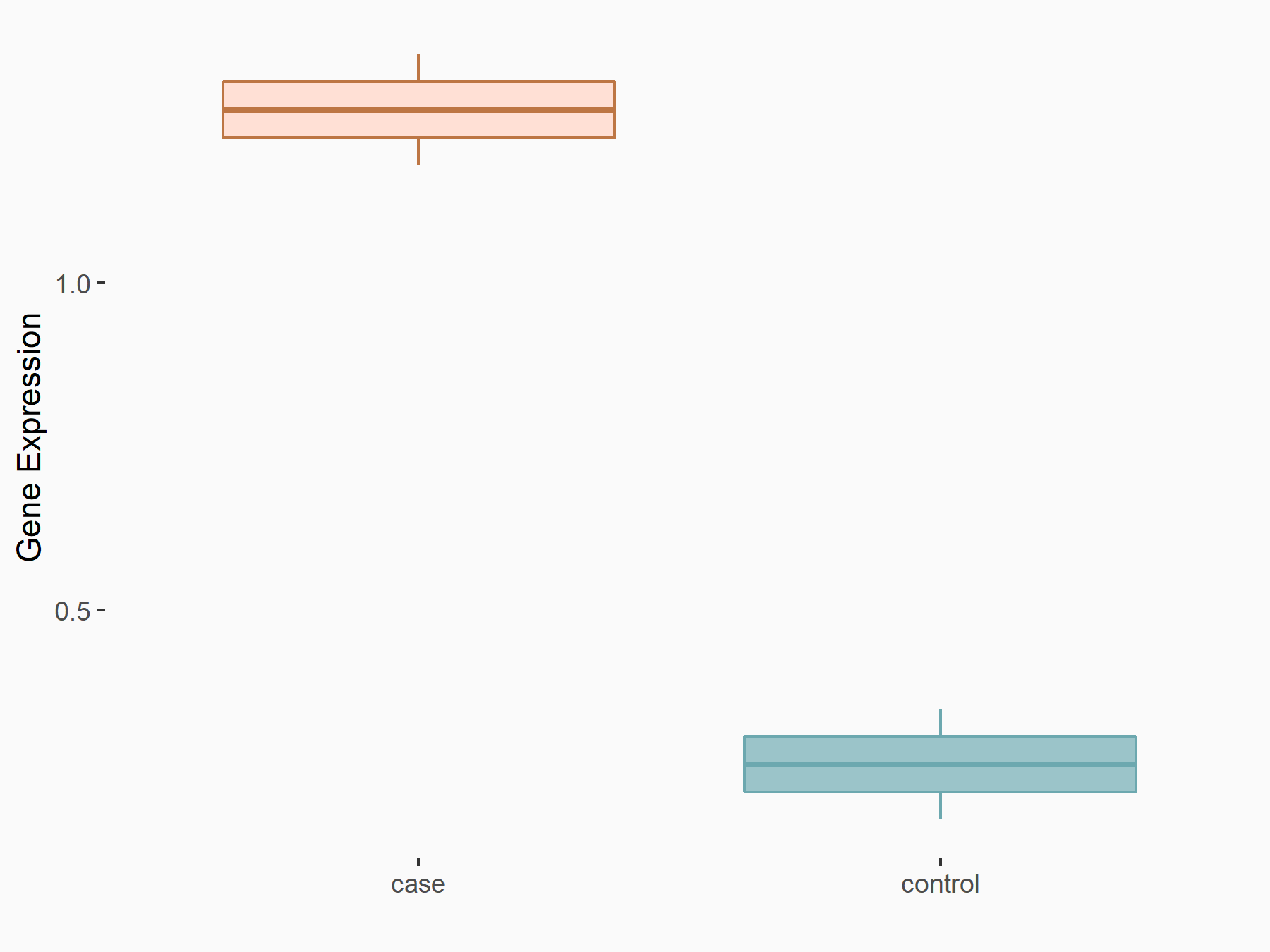
| Representative RNA-seq result indicating the expression of this target gene regulated by FTO | ||
| Cell Line | HEK293 cell line | Homo sapiens |
|
Treatment: FTO knockdown HEK293T cells
Control: HEK293T cells
|
GSE78040 | |
| Regulation |
  |
logFC: 8.43E-01 p-value: 7.94E-03 |
| More Results | Click to View More RNA-seq Results | |
| In total 1 item(s) under this regulator | ||||
| Experiment 1 Reporting the m6A Methylation Regulator of This Target Gene | [1] | |||
| Response Summary | FTO was colocalized with Matrix metalloproteinase-24 (MMP24) in spinal neurons and shown increased binding to the Mmp24 mRNA in the spinal cord after SNL. SNL promoted the m6A eraser FTO binding to the Mmp24 mRNA, which subsequently facilitated the translation of MMP24 in the spinal cord, and ultimately contributed to neuropathic pain genesis. | |||
| Target Regulation | Up regulation | |||
| Responsed Disease | Neuropathic Pain | ICD-11: 8E43.0 | ||
| In-vivo Model | Mice were anesthetized with Nembutal. The lower back was dissected until the transverse lumbar process was exposed. After the process was removed, the underneath L4 spinal nerve was ligated with a silk 6-0 thread. A slight distal location was chosen for transection around the ligation site. Subsequent layers of muscle and skin were closed. The sham groups undertook identical procedures, but without the transection or ligature of the corresponding nerve. The intraspinal injection was performed as described previously. In short, after anesthetized with Nembutal, mice underwent hemilaminectomy at the L1-L2 vertebral segments. The intraspinal injection was carried out ipsilaterally on the left side. By using a glass micropipette, each animal received two injections (5 × 10⁵ TU per injection, 0.8 mm from the midline, 0.5 mm apart in rostrocaudal axis, 0.5 mm deep) of lentivirus following the L3-L4 dorsal root entry zone after exposure of spinal cord. The tip of glass micropipette should reach a depth of lamina II-IV of the spinal cord. Finally, the dorsal muscle and skin were sutured layer by layer. | |||
Pain disorders [ICD-11: 8E43]
| In total 1 item(s) under this disease | ||||
| Experiment 1 Reporting the m6A-centered Disease Response | [1] | |||
| Response Summary | FTO was colocalized with Matrix metalloproteinase-24 (MMP24) in spinal neurons and shown increased binding to the Mmp24 mRNA in the spinal cord after SNL. SNL promoted the m6A eraser FTO binding to the Mmp24 mRNA, which subsequently facilitated the translation of MMP24 in the spinal cord, and ultimately contributed to neuropathic pain genesis. | |||
| Responsed Disease | Neuropathic Pain [ICD-11: 8E43.0] | |||
| Target Regulator | Fat mass and obesity-associated protein (FTO) | ERASER | ||
| Target Regulation | Up regulation | |||
| In-vivo Model | Mice were anesthetized with Nembutal. The lower back was dissected until the transverse lumbar process was exposed. After the process was removed, the underneath L4 spinal nerve was ligated with a silk 6-0 thread. A slight distal location was chosen for transection around the ligation site. Subsequent layers of muscle and skin were closed. The sham groups undertook identical procedures, but without the transection or ligature of the corresponding nerve. The intraspinal injection was performed as described previously. In short, after anesthetized with Nembutal, mice underwent hemilaminectomy at the L1-L2 vertebral segments. The intraspinal injection was carried out ipsilaterally on the left side. By using a glass micropipette, each animal received two injections (5 × 10⁵ TU per injection, 0.8 mm from the midline, 0.5 mm apart in rostrocaudal axis, 0.5 mm deep) of lentivirus following the L3-L4 dorsal root entry zone after exposure of spinal cord. The tip of glass micropipette should reach a depth of lamina II-IV of the spinal cord. Finally, the dorsal muscle and skin were sutured layer by layer. | |||
RNA Modification Sequencing Data Associated with the Target (ID: M6ATAR00687)
| In total 24 m6A sequence/site(s) in this target gene | |||
| mod ID: M6ASITE052253 | Click to Show/Hide the Full List | ||
| mod site | chr20:35263839-35263840:+ | [2] | |
| Sequence | GAGCTGGGCCACGCGCTGGGACTGGAGCACTCCAGCGACCC | ||
| Motif Score | 4.065041667 | ||
| Cell/Tissue List | A549 | ||
| Seq Type List | m6A-seq | ||
| Transcript ID List | ENST00000246186.8 | ||
| External Link | RMBase: m6A_site_528666 | ||
| mod ID: M6ASITE052254 | Click to Show/Hide the Full List | ||
| mod site | chr20:35267286-35267287:+ | [3] | |
| Sequence | CACTCACCATCGGAGAGGAAACACGAGCGCCAGCCCAGGCC | ||
| Motif Score | 2.20572619 | ||
| Cell/Tissue List | HeLa | ||
| Seq Type List | m6A-seq | ||
| Transcript ID List | ENST00000246186.8 | ||
| External Link | RMBase: m6A_site_528667 | ||
| mod ID: M6ASITE052255 | Click to Show/Hide the Full List | ||
| mod site | chr20:35267327-35267328:+ | [3] | |
| Sequence | CCCTCGGCCGCCCCTCGGGGACCGGCCATCCACACCAGGCA | ||
| Motif Score | 3.622404762 | ||
| Cell/Tissue List | HeLa; TREX | ||
| Seq Type List | m6A-seq; MeRIP-seq | ||
| Transcript ID List | ENST00000246186.8 | ||
| External Link | RMBase: m6A_site_528668 | ||
| mod ID: M6ASITE052256 | Click to Show/Hide the Full List | ||
| mod site | chr20:35267352-35267353:+ | [3] | |
| Sequence | CCATCCACACCAGGCACCAAACCCAACATCTGTGACGGCAA | ||
| Motif Score | 2.185083333 | ||
| Cell/Tissue List | HeLa; TREX | ||
| Seq Type List | m6A-seq; MeRIP-seq | ||
| Transcript ID List | ENST00000246186.8 | ||
| External Link | RMBase: m6A_site_528669 | ||
| mod ID: M6ASITE052257 | Click to Show/Hide the Full List | ||
| mod site | chr20:35271683-35271684:+ | [3] | |
| Sequence | GACACAGCTCTGCGCTGGGAACCTGTGGGCAAGACCTACTT | ||
| Motif Score | 2.930744048 | ||
| Cell/Tissue List | HeLa | ||
| Seq Type List | m6A-seq | ||
| Transcript ID List | ENST00000246186.8 | ||
| External Link | RMBase: m6A_site_528672 | ||
| mod ID: M6ASITE052258 | Click to Show/Hide the Full List | ||
| mod site | chr20:35271696-35271697:+ | [3] | |
| Sequence | GCTGGGAACCTGTGGGCAAGACCTACTTTTTCAAAGGCGAG | ||
| Motif Score | 2.876744048 | ||
| Cell/Tissue List | HeLa | ||
| Seq Type List | m6A-seq | ||
| Transcript ID List | ENST00000246186.8 | ||
| External Link | RMBase: m6A_site_528673 | ||
| mod ID: M6ASITE052259 | Click to Show/Hide the Full List | ||
| mod site | chr20:35271754-35271755:+ | [3] | |
| Sequence | CGAGGAGCGGCGGGCCACGGACCCTGGCTACCCTAAGCCCA | ||
| Motif Score | 3.622404762 | ||
| Cell/Tissue List | HeLa; A549 | ||
| Seq Type List | m6A-seq | ||
| Transcript ID List | ENST00000246186.8 | ||
| External Link | RMBase: m6A_site_528674 | ||
| mod ID: M6ASITE052260 | Click to Show/Hide the Full List | ||
| mod site | chr20:35274298-35274299:+ | [3] | |
| Sequence | CTATTTCTACAAGGGCCGGGACTACTGGAAGTTTGACAACC | ||
| Motif Score | 4.065041667 | ||
| Cell/Tissue List | HeLa; A549 | ||
| Seq Type List | m6A-seq | ||
| Transcript ID List | ENST00000246186.8 | ||
| External Link | RMBase: m6A_site_528675 | ||
| mod ID: M6ASITE052261 | Click to Show/Hide the Full List | ||
| mod site | chr20:35274323-35274324:+ | [3] | |
| Sequence | TGGAAGTTTGACAACCAGAAACTGAGCGTGGAGCCAGGCTA | ||
| Motif Score | 2.627720238 | ||
| Cell/Tissue List | HeLa; H1B; H1A; A549 | ||
| Seq Type List | m6A-seq | ||
| Transcript ID List | ENST00000246186.8 | ||
| External Link | RMBase: m6A_site_528676 | ||
| mod ID: M6ASITE052262 | Click to Show/Hide the Full List | ||
| mod site | chr20:35274433-35274434:+ | [3] | |
| Sequence | GCTGCCCCAGGACGACGTGGACATCATGGTGACCATCAACG | ||
| Motif Score | 3.643047619 | ||
| Cell/Tissue List | HeLa; A549; H1A; H1B; hESCs; endometrial | ||
| Seq Type List | m6A-seq; MeRIP-seq | ||
| Transcript ID List | ENST00000246186.8 | ||
| External Link | RMBase: m6A_site_528677 | ||
| mod ID: M6ASITE052263 | Click to Show/Hide the Full List | ||
| mod site | chr20:35274550-35274551:+ | [3] | |
| Sequence | CACCATCTTCCAGTTCAAGAACAAGACAGGCCCTCAGCCTG | ||
| Motif Score | 2.951386905 | ||
| Cell/Tissue List | HeLa; A549 | ||
| Seq Type List | m6A-seq; MeRIP-seq | ||
| Transcript ID List | ENST00000246186.8 | ||
| External Link | RMBase: m6A_site_528678 | ||
| mod ID: M6ASITE052264 | Click to Show/Hide the Full List | ||
| mod site | chr20:35274555-35274556:+ | [3] | |
| Sequence | TCTTCCAGTTCAAGAACAAGACAGGCCCTCAGCCTGTCACC | ||
| Motif Score | 2.897386905 | ||
| Cell/Tissue List | HeLa; A549 | ||
| Seq Type List | m6A-seq; MeRIP-seq | ||
| Transcript ID List | ENST00000246186.8 | ||
| External Link | RMBase: m6A_site_528679 | ||
| mod ID: M6ASITE052265 | Click to Show/Hide the Full List | ||
| mod site | chr20:35275348-35275349:+ | [3] | |
| Sequence | CTGCTTGAGGCTTTAGGTGAACTAGAGGTGACTGTCTTGGT | ||
| Motif Score | 3.373380952 | ||
| Cell/Tissue List | HeLa; A549 | ||
| Seq Type List | m6A-seq | ||
| Transcript ID List | ENST00000246186.8 | ||
| External Link | RMBase: m6A_site_528680 | ||
| mod ID: M6ASITE052266 | Click to Show/Hide the Full List | ||
| mod site | chr20:35275358-35275359:+ | [4] | |
| Sequence | CTTTAGGTGAACTAGAGGTGACTGTCTTGGTGATGAGGCCA | ||
| Motif Score | 3.28175 | ||
| Cell/Tissue List | A549 | ||
| Seq Type List | m6A-CLIP/IP | ||
| Transcript ID List | ENST00000246186.8 | ||
| External Link | RMBase: m6A_site_528681 | ||
| mod ID: M6ASITE052267 | Click to Show/Hide the Full List | ||
| mod site | chr20:35275408-35275409:+ | [3] | |
| Sequence | CCCTCCCCCAGGCGACAAGGACCAAGGTGCTGCTAAGGCCA | ||
| Motif Score | 3.622404762 | ||
| Cell/Tissue List | HeLa; H1A; H1B; A549 | ||
| Seq Type List | m6A-seq | ||
| Transcript ID List | ENST00000246186.8 | ||
| External Link | RMBase: m6A_site_528682 | ||
| mod ID: M6ASITE052268 | Click to Show/Hide the Full List | ||
| mod site | chr20:35275442-35275443:+ | [3] | |
| Sequence | AAGGCCACTCTAGCGCCCAGACACCCCAGTAGCTGAGCTCT | ||
| Motif Score | 2.897386905 | ||
| Cell/Tissue List | HeLa; A549; H1A; H1B | ||
| Seq Type List | m6A-seq; MeRIP-seq | ||
| Transcript ID List | ENST00000246186.8 | ||
| External Link | RMBase: m6A_site_528683 | ||
| mod ID: M6ASITE052269 | Click to Show/Hide the Full List | ||
| mod site | chr20:35276403-35276404:+ | [5] | |
| Sequence | CACCTGTCCACTCACATGAAACTCGTGTTAGGCCCTGGGAG | ||
| Motif Score | 2.627720238 | ||
| Cell/Tissue List | peripheral-blood; endometrial | ||
| Seq Type List | m6A-seq | ||
| Transcript ID List | ENST00000246186.8 | ||
| External Link | RMBase: m6A_site_528685 | ||
| mod ID: M6ASITE052270 | Click to Show/Hide the Full List | ||
| mod site | chr20:35276526-35276527:+ | [3] | |
| Sequence | TCTTGTGTCCCCATCTGTGGACCCCTCTAGGGTCTGAGATG | ||
| Motif Score | 3.622404762 | ||
| Cell/Tissue List | HeLa; H1A; H1B; fibroblasts; MM6; Huh7; Jurkat; CD4T; peripheral-blood; endometrial; NB4 | ||
| Seq Type List | m6A-seq; MeRIP-seq | ||
| Transcript ID List | ENST00000246186.8 | ||
| External Link | RMBase: m6A_site_528688 | ||
| mod ID: M6ASITE052271 | Click to Show/Hide the Full List | ||
| mod site | chr20:35276710-35276711:+ | [3] | |
| Sequence | GGCAGGCCAGCTGAGAATGAACAGGAGATGGAGGCAGGCAG | ||
| Motif Score | 2.951386905 | ||
| Cell/Tissue List | HeLa; HEK293T; H1A; H1B; MM6; peripheral-blood; endometrial | ||
| Seq Type List | m6A-seq; MeRIP-seq | ||
| Transcript ID List | ENST00000246186.8 | ||
| External Link | RMBase: m6A_site_528693 | ||
| mod ID: M6ASITE052272 | Click to Show/Hide the Full List | ||
| mod site | chr20:35276841-35276842:+ | [3] | |
| Sequence | TGTTTGTCCATCACCCCAGGACAGGGCAGACAGAGGGGCAA | ||
| Motif Score | 3.643047619 | ||
| Cell/Tissue List | HeLa; HEK293T; kidney; H1A; H1B; fibroblasts; A549; MM6; Jurkat; peripheral-blood; HEK293A-TOA; TREX; endometrial | ||
| Seq Type List | m6A-seq; MeRIP-seq; m6A-REF-seq | ||
| Transcript ID List | ENST00000246186.8 | ||
| External Link | RMBase: m6A_site_528696 | ||
| mod ID: M6ASITE052273 | Click to Show/Hide the Full List | ||
| mod site | chr20:35276850-35276851:+ | [3] | |
| Sequence | ATCACCCCAGGACAGGGCAGACAGAGGGGCAAAGCACTGGG | ||
| Motif Score | 2.897386905 | ||
| Cell/Tissue List | HeLa; HEK293T; H1A; H1B; fibroblasts; A549; MM6; Jurkat; peripheral-blood; HEK293A-TOA; TREX; endometrial | ||
| Seq Type List | m6A-seq; MeRIP-seq | ||
| Transcript ID List | ENST00000246186.8 | ||
| External Link | RMBase: m6A_site_528697 | ||
| mod ID: M6ASITE052274 | Click to Show/Hide the Full List | ||
| mod site | chr20:35276905-35276906:+ | [3] | |
| Sequence | GCTTCCCCTCAGCCTGGGGGACATCACAGCATTTCAGTGTC | ||
| Motif Score | 3.643047619 | ||
| Cell/Tissue List | HeLa; HEK293T; H1A; H1B; hNPCs; fibroblasts; A549; MM6; Jurkat; peripheral-blood; HEK293A-TOA; TREX; endometrial | ||
| Seq Type List | m6A-seq; MeRIP-seq | ||
| Transcript ID List | ENST00000246186.8 | ||
| External Link | RMBase: m6A_site_528698 | ||
| mod ID: M6ASITE052275 | Click to Show/Hide the Full List | ||
| mod site | chr20:35276939-35276940:+ | [3] | |
| Sequence | CAGTGTCAGTCACATTTTAAACTGATCAGCCTTTGTATAAT | ||
| Motif Score | 2.627720238 | ||
| Cell/Tissue List | HeLa; HEK293T; H1A; H1B; hESCs; HEK293A-TOA | ||
| Seq Type List | m6A-seq; MeRIP-seq | ||
| Transcript ID List | ENST00000246186.8 | ||
| External Link | RMBase: m6A_site_528701 | ||
| mod ID: M6ASITE052276 | Click to Show/Hide the Full List | ||
| mod site | chr20:35276985-35276986:+ | [3] | |
| Sequence | TTAAATCATTTCTAAATAAAACAGAAATACAGAGTGTGTCA | ||
| Motif Score | 2.20572619 | ||
| Cell/Tissue List | HeLa; HEK293T; hESCs; HEK293A-TOA | ||
| Seq Type List | m6A-seq; MeRIP-seq | ||
| Transcript ID List | ENST00000246186.8 | ||
| External Link | RMBase: m6A_site_528704 | ||
References