m6A Target Gene Information
General Information of the m6A Target Gene (ID: M6ATAR00621)
Full List of m6A Methylation Regulator of This Target Gene and Corresponding Disease/Drug Response(s)
PKC-eta
can be regulated by the following regulator(s), and cause disease/drug response(s). You can browse detail information of regulator(s) or disease/drug response(s).
Browse Regulator
Browse Disease
Methyltransferase-like 3 (METTL3) [WRITER]
| Representative RNA-seq result indicating the expression of this target gene regulated by METTL3 | ||
| Cell Line | Caco-2 cell line | Homo sapiens |
|
Treatment: shMETTL3 Caco-2 cells
Control: shNTC Caco-2 cells
|
GSE167075 | |
| Regulation |
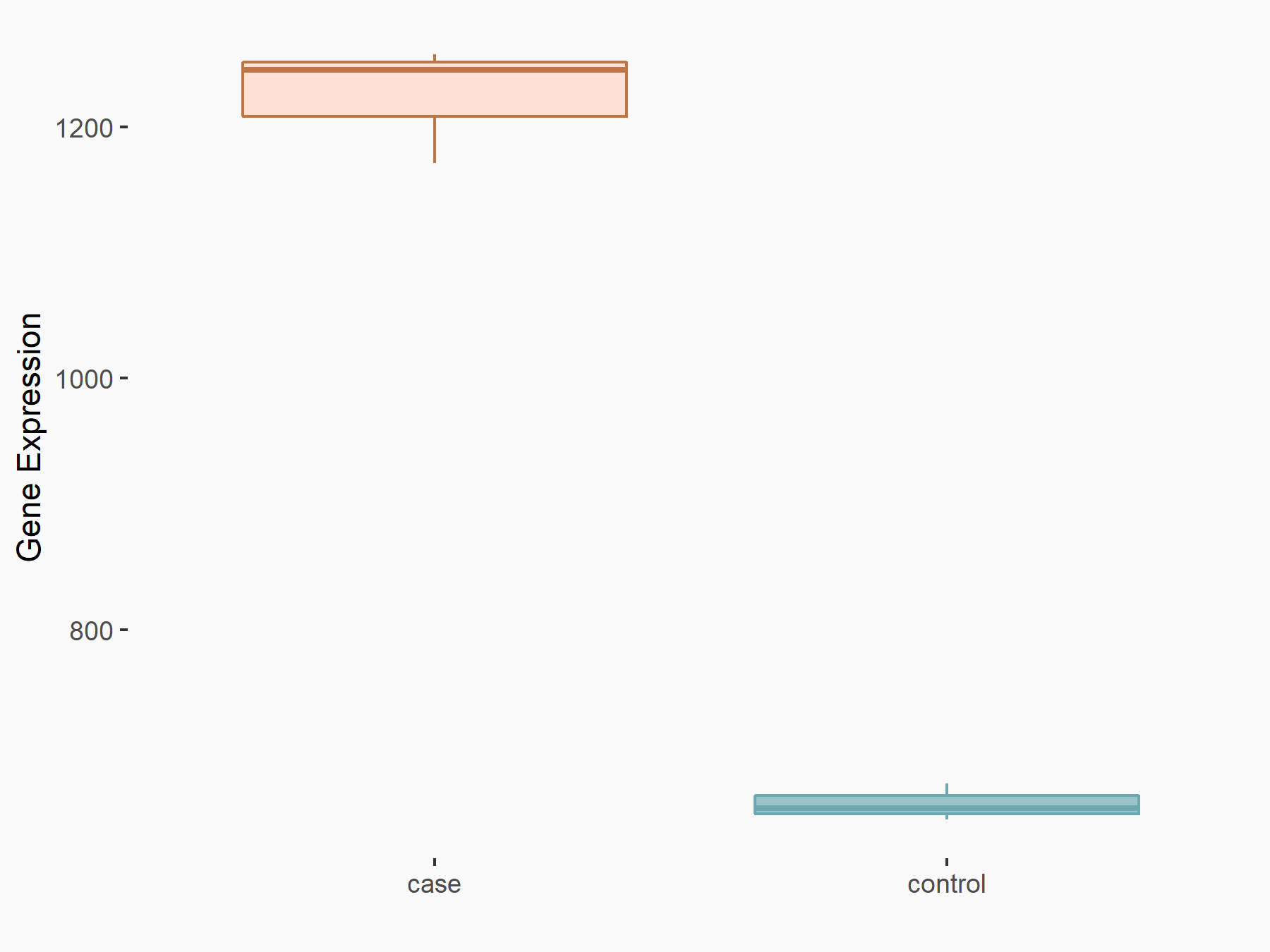 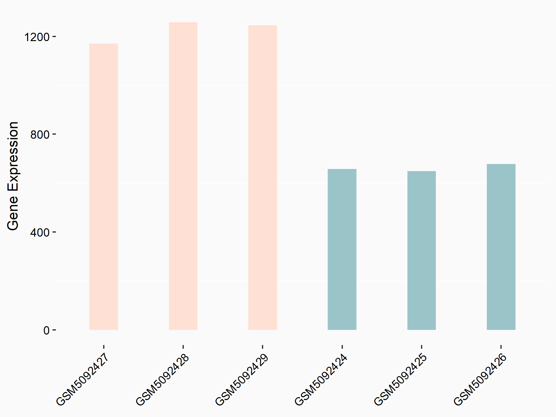 |
logFC: 8.88E-01 p-value: 2.37E-29 |
| More Results | Click to View More RNA-seq Results | |
| In total 1 item(s) under this regulator | ||||
| Experiment 1 Reporting the m6A Methylation Regulator of This Target Gene | [1] | |||
| Response Summary | Specific depletion of METTL3 in pericytes suppressed diabetes-induced pericyte dysfunction and Microvascular complication in vivo. METTL3 overexpression impaired pericyte function by repressing Protein kinase C eta type (PKC-eta), FAT4, and PDGFRA expression, which was mediated by YTHDF2-dependent mRNA decay. | |||
| Target Regulation | Down regulation | |||
| Responsed Disease | Diseases of arteries or arterioles | ICD-11: BD5Y | ||
| In-vitro Model | ACBRI-183 (Human retinal pericytes (ACBRI-183) was obtained from Cell Systems Corp. (CSC, USA)) | |||
| In-vivo Model | Mettl3 floxed mice were purchased from GemPharmatech Co. Ltd (Nanjing, China). Pdgfr-Beta-Cre mice were purchased from Beijing Biocytogen Co. Ltd (Beijing, China) generated on C57BL/6J background. Mettl3 flox/flox mice were crossed with Pdgfr-Beta-Cre mice to generate pericyte-specific Mettl3 knockout mice. All mice were bred under the specific-pathogen free condition with free access to diet and water or their nursing mothers with alternating 12/12 light-dark cycle (lights on at 08:00 and off at 20:00). | |||
Diseases of arteries or arterioles [ICD-11: BD5Y]
| In total 1 item(s) under this disease | ||||
| Experiment 1 Reporting the m6A-centered Disease Response | [1] | |||
| Response Summary | Specific depletion of METTL3 in pericytes suppressed diabetes-induced pericyte dysfunction and Microvascular complication in vivo. METTL3 overexpression impaired pericyte function by repressing Protein kinase C eta type (PKC-eta), FAT4, and PDGFRA expression, which was mediated by YTHDF2-dependent mRNA decay. | |||
| Responsed Disease | Diseases of arteries or arterioles [ICD-11: BD5Y] | |||
| Target Regulator | Methyltransferase-like 3 (METTL3) | WRITER | ||
| Target Regulation | Down regulation | |||
| In-vitro Model | ACBRI-183 (Human retinal pericytes (ACBRI-183) was obtained from Cell Systems Corp. (CSC, USA)) | |||
| In-vivo Model | Mettl3 floxed mice were purchased from GemPharmatech Co. Ltd (Nanjing, China). Pdgfr-Beta-Cre mice were purchased from Beijing Biocytogen Co. Ltd (Beijing, China) generated on C57BL/6J background. Mettl3 flox/flox mice were crossed with Pdgfr-Beta-Cre mice to generate pericyte-specific Mettl3 knockout mice. All mice were bred under the specific-pathogen free condition with free access to diet and water or their nursing mothers with alternating 12/12 light-dark cycle (lights on at 08:00 and off at 20:00). | |||
RNA Modification Sequencing Data Associated with the Target (ID: M6ATAR00621)
| In total 48 m6A sequence/site(s) in this target gene | |||
| mod ID: M6ASITE019879 | Click to Show/Hide the Full List | ||
| mod site | chr14:61281162-61281163:+ | [2] | |
| Sequence | GTCATGGCGGCCGCGCGGGGACCGACGCGCGGGCTGACCGA | ||
| Motif Score | 3.622404762 | ||
| Cell/Tissue List | H1A; H1B | ||
| Seq Type List | m6A-seq | ||
| Transcript ID List | ENST00000555185.5; ENST00000556778.5; ENST00000555906.5; ENST00000557294.5; ENST00000555542.5 | ||
| External Link | RMBase: m6A_site_250303 | ||
| mod ID: M6ASITE019880 | Click to Show/Hide the Full List | ||
| mod site | chr14:61281235-61281236:+ | [2] | |
| Sequence | CGCGCCGCGCAAGCCCGGGGACTGCTCGCGGGTCGGGTTTC | ||
| Motif Score | 4.065041667 | ||
| Cell/Tissue List | H1A; H1B | ||
| Seq Type List | m6A-seq | ||
| Transcript ID List | ENST00000555542.5; ENST00000557294.5; ENST00000556778.5; ENST00000555185.5; ENST00000554835.1; ENST00000555906.5 | ||
| External Link | RMBase: m6A_site_250304 | ||
| mod ID: M6ASITE019881 | Click to Show/Hide the Full List | ||
| mod site | chr14:61322040-61322041:+ | [3] | |
| Sequence | GAGGGCTGGCCTGAGACGGGACTCCCGGTTCTCCCGCTGCG | ||
| Motif Score | 4.065041667 | ||
| Cell/Tissue List | HeLa; HEK293T; Jurkat; CD4T; endometrial | ||
| Seq Type List | m6A-seq; MeRIP-seq | ||
| Transcript ID List | ENST00000555906.5; ENST00000556778.5; ENST00000557294.5; ENST00000332981.10; ENST00000555542.5; ENST00000555185.5 | ||
| External Link | RMBase: m6A_site_250313 | ||
| mod ID: M6ASITE019882 | Click to Show/Hide the Full List | ||
| mod site | chr14:61322225-61322226:+ | [4] | |
| Sequence | GAAGGGCCACCAGCTGCTGGACCCCTATCTGACGGTGAGCG | ||
| Motif Score | 3.622404762 | ||
| Cell/Tissue List | Jurkat; CD4T; HEK293T; endometrial | ||
| Seq Type List | m6A-seq | ||
| Transcript ID List | ENST00000555542.5; ENST00000332981.10; ENST00000555906.5; ENST00000556778.5; ENST00000555185.5; ENST00000557294.5 | ||
| External Link | RMBase: m6A_site_250314 | ||
| mod ID: M6ASITE019883 | Click to Show/Hide the Full List | ||
| mod site | chr14:61322249-61322250:+ | [4] | |
| Sequence | CTATCTGACGGTGAGCGTGGACCAGGTGCGCGTGGGCCAGA | ||
| Motif Score | 3.622404762 | ||
| Cell/Tissue List | Jurkat; CD4T; HEK293T; endometrial | ||
| Seq Type List | m6A-seq | ||
| Transcript ID List | ENST00000555906.5; ENST00000555542.5; ENST00000332981.10; ENST00000555185.5; ENST00000557294.5; ENST00000556778.5 | ||
| External Link | RMBase: m6A_site_250315 | ||
| mod ID: M6ASITE019884 | Click to Show/Hide the Full List | ||
| mod site | chr14:61322269-61322270:+ | [4] | |
| Sequence | ACCAGGTGCGCGTGGGCCAGACCAGCACCAAGCAGAAGACC | ||
| Motif Score | 2.876744048 | ||
| Cell/Tissue List | Jurkat; CD4T; HEK293T; endometrial | ||
| Seq Type List | m6A-seq | ||
| Transcript ID List | ENST00000556778.5; ENST00000555542.5; ENST00000555906.5; ENST00000332981.10; ENST00000557294.5; ENST00000555185.5 | ||
| External Link | RMBase: m6A_site_250316 | ||
| mod ID: M6ASITE019885 | Click to Show/Hide the Full List | ||
| mod site | chr14:61322287-61322288:+ | [4] | |
| Sequence | AGACCAGCACCAAGCAGAAGACCAACAAACCCACGTACAAC | ||
| Motif Score | 2.876744048 | ||
| Cell/Tissue List | Jurkat; CD4T; HEK293T; endometrial | ||
| Seq Type List | m6A-seq | ||
| Transcript ID List | ENST00000555906.5; ENST00000555185.5; ENST00000556778.5; ENST00000332981.10; ENST00000555542.5; ENST00000557294.5 | ||
| External Link | RMBase: m6A_site_250317 | ||
| mod ID: M6ASITE019886 | Click to Show/Hide the Full List | ||
| mod site | chr14:61322295-61322296:+ | [4] | |
| Sequence | ACCAAGCAGAAGACCAACAAACCCACGTACAACGAGGAGTT | ||
| Motif Score | 2.185083333 | ||
| Cell/Tissue List | Jurkat; CD4T; HEK293T; endometrial | ||
| Seq Type List | m6A-seq | ||
| Transcript ID List | ENST00000556778.5; ENST00000555542.5; ENST00000555185.5; ENST00000557294.5; ENST00000332981.10; ENST00000555906.5 | ||
| External Link | RMBase: m6A_site_250318 | ||
| mod ID: M6ASITE019887 | Click to Show/Hide the Full List | ||
| mod site | chr14:61322303-61322304:+ | [5] | |
| Sequence | GAAGACCAACAAACCCACGTACAACGAGGAGTTTTGCGCTA | ||
| Motif Score | 2.856142857 | ||
| Cell/Tissue List | hESC-HEK293T | ||
| Seq Type List | MAZTER-seq | ||
| Transcript ID List | ENST00000332981.10; ENST00000555906.5; ENST00000555542.5; ENST00000556778.5; ENST00000557294.5; ENST00000555185.5 | ||
| External Link | RMBase: m6A_site_250319 | ||
| mod ID: M6ASITE019888 | Click to Show/Hide the Full List | ||
| mod site | chr14:61322447-61322448:+ | [6] | |
| Sequence | GCGCACGACCGGCGCCTCGGACACCTTCGAGGGTTGGGTGA | ||
| Motif Score | 3.643047619 | ||
| Cell/Tissue List | CD34; hESC-HEK293T | ||
| Seq Type List | m6A-seq; MAZTER-seq | ||
| Transcript ID List | ENST00000556778.5; ENST00000332981.10; ENST00000555906.5; ENST00000555542.5; ENST00000555185.5; ENST00000557294.5 | ||
| External Link | RMBase: m6A_site_250320 | ||
| mod ID: M6ASITE019889 | Click to Show/Hide the Full List | ||
| mod site | chr14:61322674-61322675:+ | [6] | |
| Sequence | TCTCCAGGAAGCCGGCCTGGACACGATCCCCTTTCAGGACA | ||
| Motif Score | 3.643047619 | ||
| Cell/Tissue List | CD34 | ||
| Seq Type List | m6A-seq | ||
| Transcript ID List | ENST00000332981.10; ENST00000555906.5; ENST00000556778.5; ENST00000555542.5; ENST00000555082.5; ENST00000555185.5 | ||
| External Link | RMBase: m6A_site_250321 | ||
| mod ID: M6ASITE019890 | Click to Show/Hide the Full List | ||
| mod site | chr14:61322692-61322693:+ | [6] | |
| Sequence | GGACACGATCCCCTTTCAGGACAGCGGCTTGGCCTAACGGG | ||
| Motif Score | 3.643047619 | ||
| Cell/Tissue List | CD34 | ||
| Seq Type List | m6A-seq | ||
| Transcript ID List | ENST00000332981.10; ENST00000555185.5; ENST00000555542.5; ENST00000555082.5; ENST00000556778.5; ENST00000555906.5 | ||
| External Link | RMBase: m6A_site_250322 | ||
| mod ID: M6ASITE019891 | Click to Show/Hide the Full List | ||
| mod site | chr14:61443138-61443139:+ | [5] | |
| Sequence | CAGAGAGACCGGATCTTCAAACATTTTACCAGGAAGCGCCA | ||
| Motif Score | 2.20572619 | ||
| Cell/Tissue List | hESC-HEK293T | ||
| Seq Type List | MAZTER-seq | ||
| Transcript ID List | ENST00000553265.5; ENST00000553726.5; ENST00000553831.5; ENST00000555542.5; ENST00000332981.10; ENST00000556164.5; ENST00000555082.5; ENST00000557473.1; ENST00000557585.5; ENST00000556778.5; ENST00000555906.5; ENST00000553830.1; ENST00000555185.5 | ||
| External Link | RMBase: m6A_site_250323 | ||
| mod ID: M6ASITE019892 | Click to Show/Hide the Full List | ||
| mod site | chr14:61450907-61450908:+ | [5] | |
| Sequence | TCCACAACTACAAAGTGCCAACATTCTGCGATCACTGTGGC | ||
| Motif Score | 2.173910714 | ||
| Cell/Tissue List | hESC-HEK293T | ||
| Seq Type List | MAZTER-seq | ||
| Transcript ID List | ENST00000332981.10; ENST00000555082.5; ENST00000556778.5; ENST00000555185.5; ENST00000553726.5; ENST00000557473.1; ENST00000557585.5 | ||
| External Link | RMBase: m6A_site_250324 | ||
| mod ID: M6ASITE019893 | Click to Show/Hide the Full List | ||
| mod site | chr14:61450951-61450952:+ | [5] | |
| Sequence | CTGCTCTGGGGAATAATGCGACAAGGACTTCAGTGTAAAAG | ||
| Motif Score | 2.865571429 | ||
| Cell/Tissue List | hESC-HEK293T | ||
| Seq Type List | MAZTER-seq | ||
| Transcript ID List | ENST00000557585.5; ENST00000556778.5; ENST00000555082.5; ENST00000553726.5; ENST00000555185.5; ENST00000332981.10 | ||
| External Link | RMBase: m6A_site_250325 | ||
| mod ID: M6ASITE019894 | Click to Show/Hide the Full List | ||
| mod site | chr14:61453295-61453296:+ | [6] | |
| Sequence | TGTGGGGTAAATGCGGTGGAACTTGCCAAGACCCTGGCAGG | ||
| Motif Score | 3.373380952 | ||
| Cell/Tissue List | CD34 | ||
| Seq Type List | m6A-seq | ||
| Transcript ID List | ENST00000553726.5; ENST00000332981.10; ENST00000555082.5; ENST00000555185.5; ENST00000557585.5; ENST00000553889.5 | ||
| External Link | RMBase: m6A_site_250326 | ||
| mod ID: M6ASITE019895 | Click to Show/Hide the Full List | ||
| mod site | chr14:61453305-61453306:+ | [6] | |
| Sequence | ATGCGGTGGAACTTGCCAAGACCCTGGCAGGGATGGGTCTC | ||
| Motif Score | 2.876744048 | ||
| Cell/Tissue List | CD34 | ||
| Seq Type List | m6A-seq | ||
| Transcript ID List | ENST00000555185.5; ENST00000332981.10; ENST00000557585.5; ENST00000553726.5; ENST00000553889.5; ENST00000555082.5 | ||
| External Link | RMBase: m6A_site_250327 | ||
| mod ID: M6ASITE019896 | Click to Show/Hide the Full List | ||
| mod site | chr14:61456846-61456847:+ | [6] | |
| Sequence | GGTGGCGCTCGTGGAATGGGACCTTCCCGTGGAAAGCCACC | ||
| Motif Score | 3.622404762 | ||
| Cell/Tissue List | CD34 | ||
| Seq Type List | m6A-seq | ||
| Transcript ID List | ENST00000557585.5; ENST00000555185.5; ENST00000555082.5; ENST00000553726.5; ENST00000557559.1; ENST00000553889.5; ENST00000332981.10 | ||
| External Link | RMBase: m6A_site_250328 | ||
| mod ID: M6ASITE019897 | Click to Show/Hide the Full List | ||
| mod site | chr14:61457031-61457032:+ | [7] | |
| Sequence | AGGGAGTTTCGAGTGGCAGAACCGATCATCTGTCAAACTGA | ||
| Motif Score | 2.930744048 | ||
| Cell/Tissue List | endometrial | ||
| Seq Type List | m6A-seq | ||
| Transcript ID List | ENST00000553726.5; ENST00000557585.5; ENST00000555082.5; ENST00000332981.10; ENST00000553889.5; ENST00000557559.1; ENST00000555185.5 | ||
| External Link | RMBase: m6A_site_250329 | ||
| mod ID: M6ASITE019898 | Click to Show/Hide the Full List | ||
| mod site | chr14:61457047-61457048:+ | [7] | |
| Sequence | CAGAACCGATCATCTGTCAAACTGAGTTGATCTTTCCCCCA | ||
| Motif Score | 2.627720238 | ||
| Cell/Tissue List | endometrial | ||
| Seq Type List | m6A-seq | ||
| Transcript ID List | ENST00000555082.5; ENST00000557585.5; ENST00000332981.10; ENST00000553889.5; ENST00000557559.1; ENST00000555185.5; ENST00000553726.5 | ||
| External Link | RMBase: m6A_site_250330 | ||
| mod ID: M6ASITE019899 | Click to Show/Hide the Full List | ||
| mod site | chr14:61457177-61457178:+ | [7] | |
| Sequence | GTGTTCTTACTCTTTCAGAAACTCGTTTCCAGATCGACCCT | ||
| Motif Score | 2.627720238 | ||
| Cell/Tissue List | endometrial | ||
| Seq Type List | m6A-seq | ||
| Transcript ID List | ENST00000553889.5; ENST00000557585.5; ENST00000553726.5; ENST00000332981.10; ENST00000555185.5; ENST00000557559.1; ENST00000555082.5 | ||
| External Link | RMBase: m6A_site_250331 | ||
| mod ID: M6ASITE019900 | Click to Show/Hide the Full List | ||
| mod site | chr14:61457529-61457530:+ | [6] | |
| Sequence | TGCTTGCAAGAGTAAAAGAAACAGGAGACCTCTATGCTGTG | ||
| Motif Score | 2.20572619 | ||
| Cell/Tissue List | CD34; endometrial | ||
| Seq Type List | m6A-seq | ||
| Transcript ID List | ENST00000332981.10; ENST00000555185.5; ENST00000553889.5; ENST00000557585.5; ENST00000555604.1; ENST00000555082.5; ENST00000557559.1 | ||
| External Link | RMBase: m6A_site_250332 | ||
| mod ID: M6ASITE019901 | Click to Show/Hide the Full List | ||
| mod site | chr14:61457536-61457537:+ | [6] | |
| Sequence | AAGAGTAAAAGAAACAGGAGACCTCTATGCTGTGAAGGTGC | ||
| Motif Score | 2.876744048 | ||
| Cell/Tissue List | CD34; endometrial | ||
| Seq Type List | m6A-seq | ||
| Transcript ID List | ENST00000557585.5; ENST00000557559.1; ENST00000555185.5; ENST00000553889.5; ENST00000332981.10; ENST00000555604.1; ENST00000555082.5 | ||
| External Link | RMBase: m6A_site_250333 | ||
| mod ID: M6ASITE019902 | Click to Show/Hide the Full List | ||
| mod site | chr14:61529088-61529089:+ | [5] | |
| Sequence | TTTCAGAGATCTGAAACTGGACAATGTCCTGTTGGACCACG | ||
| Motif Score | 3.643047619 | ||
| Cell/Tissue List | hESC-HEK293T | ||
| Seq Type List | MAZTER-seq | ||
| Transcript ID List | ENST00000555233.5; ENST00000536400.5; ENST00000555628.5; ENST00000555185.5; ENST00000557599.5; ENST00000640011.1; ENST00000555082.5; ENST00000332981.10; ENST00000555382.5; ENST00000553846.1; ENST00000555110.1 | ||
| External Link | RMBase: m6A_site_250334 | ||
| mod ID: M6ASITE019903 | Click to Show/Hide the Full List | ||
| mod site | chr14:61543388-61543389:+ | [6] | |
| Sequence | GGGCCACAGCAGGAAGGGGAACATAAAATGAGTCCCTGAGT | ||
| Motif Score | 2.951386905 | ||
| Cell/Tissue List | CD34 | ||
| Seq Type List | m6A-seq | ||
| Transcript ID List | ENST00000555082.5; ENST00000556245.1; ENST00000556347.1; ENST00000555628.5; ENST00000557599.5; ENST00000536400.5; ENST00000555382.5; ENST00000332981.10; ENST00000553846.1 | ||
| External Link | RMBase: m6A_site_250335 | ||
| mod ID: M6ASITE019904 | Click to Show/Hide the Full List | ||
| mod site | chr14:61544660-61544661:+ | [6] | |
| Sequence | CAGCCTCCCAGATTTGGCAGACTGTAGGATCTCAGTAAGTG | ||
| Motif Score | 3.319380952 | ||
| Cell/Tissue List | CD34 | ||
| Seq Type List | m6A-seq | ||
| Transcript ID List | ENST00000556347.1; ENST00000556245.1; ENST00000536400.5; ENST00000555082.5; ENST00000332981.10; ENST00000555628.5; ENST00000555382.5; ENST00000557599.5; ENST00000553846.1 | ||
| External Link | RMBase: m6A_site_250336 | ||
| mod ID: M6ASITE019905 | Click to Show/Hide the Full List | ||
| mod site | chr14:61547755-61547756:+ | [6] | |
| Sequence | CACTCAGTTCATGACCAAGAACCCCACCATGCGCTTGGGCA | ||
| Motif Score | 2.930744048 | ||
| Cell/Tissue List | CD34 | ||
| Seq Type List | m6A-seq | ||
| Transcript ID List | ENST00000553846.1; ENST00000557599.5; ENST00000555628.5; ENST00000555082.5; ENST00000332981.10; ENST00000556245.1; ENST00000555382.5; ENST00000536400.5; ENST00000556347.1 | ||
| External Link | RMBase: m6A_site_250337 | ||
| mod ID: M6ASITE019906 | Click to Show/Hide the Full List | ||
| mod site | chr14:61547810-61547811:+ | [6] | |
| Sequence | GGCGAGCACGCCATCTTGAGACATCCTTTTTTTAAGGAAAT | ||
| Motif Score | 2.897386905 | ||
| Cell/Tissue List | CD34 | ||
| Seq Type List | m6A-seq | ||
| Transcript ID List | ENST00000555628.5; ENST00000555082.5; ENST00000556245.1; ENST00000553846.1; ENST00000556347.1; ENST00000557599.5; ENST00000332981.10; ENST00000555382.5; ENST00000536400.5 | ||
| External Link | RMBase: m6A_site_250338 | ||
| mod ID: M6ASITE019907 | Click to Show/Hide the Full List | ||
| mod site | chr14:61547848-61547849:+ | [6] | |
| Sequence | AATCGACTGGGCCCAGCTGAACCATCGCCAAATAGAACCGC | ||
| Motif Score | 2.930744048 | ||
| Cell/Tissue List | CD34 | ||
| Seq Type List | m6A-seq | ||
| Transcript ID List | ENST00000332981.10; ENST00000555382.5; ENST00000557599.5; ENST00000556245.1; ENST00000555082.5; ENST00000536400.5; ENST00000556347.1; ENST00000555628.5 | ||
| External Link | RMBase: m6A_site_250339 | ||
| mod ID: M6ASITE019908 | Click to Show/Hide the Full List | ||
| mod site | chr14:61547864-61547865:+ | [6] | |
| Sequence | CTGAACCATCGCCAAATAGAACCGCCTTTCAGACCCAGAAT | ||
| Motif Score | 2.930744048 | ||
| Cell/Tissue List | CD34; HEK293T | ||
| Seq Type List | m6A-seq; MeRIP-seq | ||
| Transcript ID List | ENST00000332981.10; ENST00000536400.5; ENST00000555382.5; ENST00000557599.5; ENST00000556347.1; ENST00000556245.1; ENST00000555082.5 | ||
| External Link | RMBase: m6A_site_250340 | ||
| mod ID: M6ASITE019909 | Click to Show/Hide the Full List | ||
| mod site | chr14:61547876-61547877:+ | [6] | |
| Sequence | CAAATAGAACCGCCTTTCAGACCCAGAATCGTAAGTGTCCC | ||
| Motif Score | 2.876744048 | ||
| Cell/Tissue List | CD34; HEK293T | ||
| Seq Type List | m6A-seq; MeRIP-seq | ||
| Transcript ID List | ENST00000556245.1; ENST00000332981.10; ENST00000536400.5; ENST00000555082.5; ENST00000555382.5; ENST00000556347.1; ENST00000557599.5 | ||
| External Link | RMBase: m6A_site_250341 | ||
| mod ID: M6ASITE019910 | Click to Show/Hide the Full List | ||
| mod site | chr14:61549761-61549762:+ | [6] | |
| Sequence | TTAACTCCAATTGATGAGGGACATCTTCCAATGATTAACCA | ||
| Motif Score | 3.643047619 | ||
| Cell/Tissue List | CD34; HEK293T; hESC-HEK293T; A549; LCLs; CD8T; TIME | ||
| Seq Type List | m6A-seq; MeRIP-seq; MAZTER-seq; m6A-CLIP/IP | ||
| Transcript ID List | ENST00000332981.10; ENST00000555082.5; ENST00000557599.5; ENST00000556245.1; ENST00000556347.1; ENST00000536400.5 | ||
| External Link | RMBase: m6A_site_250342 | ||
| mod ID: M6ASITE019911 | Click to Show/Hide the Full List | ||
| mod site | chr14:61549796-61549797:+ | [6] | |
| Sequence | TAACCAGGATGAGTTTAGAAACTTTTCCTATGTGTCTCCAG | ||
| Motif Score | 2.627720238 | ||
| Cell/Tissue List | CD34; HEK293T; U2OS; A549; GM12878; LCLs; TIME | ||
| Seq Type List | m6A-seq; MeRIP-seq | ||
| Transcript ID List | ENST00000556347.1; ENST00000555082.5; ENST00000536400.5; ENST00000556245.1; ENST00000557599.5; ENST00000332981.10 | ||
| External Link | RMBase: m6A_site_250343 | ||
| mod ID: M6ASITE019912 | Click to Show/Hide the Full List | ||
| mod site | chr14:61549862-61549863:+ | [8] | |
| Sequence | GTGAGAGAGAGGGCACGAGAACCCAAAGGGAATAGAGATTC | ||
| Motif Score | 2.930744048 | ||
| Cell/Tissue List | HeLa; CD34; HEK293T; A549; U2OS; GM12878; LCLs; Jurkat; HEK293A-TOA; iSLK; MSC; TIME | ||
| Seq Type List | m6A-seq; MeRIP-seq | ||
| Transcript ID List | ENST00000536400.5; ENST00000555082.5; ENST00000556347.1; ENST00000556245.1; ENST00000332981.10 | ||
| External Link | RMBase: m6A_site_250344 | ||
| mod ID: M6ASITE019913 | Click to Show/Hide the Full List | ||
| mod site | chr14:61549904-61549905:+ | [8] | |
| Sequence | CCAGGAATTTCCTCTATGGGACCTTCCCAGCATCAGCCTTA | ||
| Motif Score | 3.622404762 | ||
| Cell/Tissue List | HeLa; CD34; HEK293T; A549; U2OS; GM12878; LCLs; Huh7; Jurkat; HEK293A-TOA; iSLK; MSC; TIME; endometrial | ||
| Seq Type List | m6A-seq; MeRIP-seq | ||
| Transcript ID List | ENST00000332981.10; ENST00000555082.5; ENST00000536400.5; ENST00000556245.1; ENST00000556347.1 | ||
| External Link | RMBase: m6A_site_250345 | ||
| mod ID: M6ASITE019914 | Click to Show/Hide the Full List | ||
| mod site | chr14:61549927-61549928:+ | [8] | |
| Sequence | TTCCCAGCATCAGCCTTAGAACAAGAACCTTACCTTCAAGG | ||
| Motif Score | 2.951386905 | ||
| Cell/Tissue List | HeLa; CD34; HEK293T; A549; hESC-HEK293T; U2OS; GM12878; LCLs; Huh7; Jurkat; HEK293A-TOA; iSLK; MSC; TIME; endometrial | ||
| Seq Type List | m6A-seq; MeRIP-seq; MAZTER-seq | ||
| Transcript ID List | ENST00000332981.10; ENST00000556245.1; ENST00000556347.1; ENST00000536400.5; ENST00000555082.5 | ||
| External Link | RMBase: m6A_site_250346 | ||
| mod ID: M6ASITE019915 | Click to Show/Hide the Full List | ||
| mod site | chr14:61549933-61549934:+ | [8] | |
| Sequence | GCATCAGCCTTAGAACAAGAACCTTACCTTCAAGGAGCAAG | ||
| Motif Score | 2.930744048 | ||
| Cell/Tissue List | HeLa; CD34; HEK293T; A549; U2OS; H1A; H1B; GM12878; LCLs; Huh7; Jurkat; HEK293A-TOA; iSLK; MSC; TIME; endometrial | ||
| Seq Type List | m6A-seq; MeRIP-seq | ||
| Transcript ID List | ENST00000536400.5; ENST00000556245.1; ENST00000332981.10; ENST00000555082.5; ENST00000556347.1 | ||
| External Link | RMBase: m6A_site_250347 | ||
| mod ID: M6ASITE019916 | Click to Show/Hide the Full List | ||
| mod site | chr14:61549960-61549961:+ | [8] | |
| Sequence | CTTCAAGGAGCAAGTGAAGAACTCTGTGAAGGATGGAACTT | ||
| Motif Score | 3.373380952 | ||
| Cell/Tissue List | HeLa; CD34; HEK293T; A549; U2OS; H1A; H1B; GM12878; LCLs; Huh7; Jurkat; HEK293A-TOA; iSLK; MSC; TIME; endometrial | ||
| Seq Type List | m6A-seq; MeRIP-seq | ||
| Transcript ID List | ENST00000556347.1; ENST00000556245.1; ENST00000332981.10; ENST00000555082.5 | ||
| External Link | RMBase: m6A_site_250348 | ||
| mod ID: M6ASITE019917 | Click to Show/Hide the Full List | ||
| mod site | chr14:61549977-61549978:+ | [8] | |
| Sequence | AGAACTCTGTGAAGGATGGAACTTTCAGATATCAACTATTT | ||
| Motif Score | 3.373380952 | ||
| Cell/Tissue List | HeLa; CD34; HEK293T; A549; U2OS; H1A; H1B; GM12878; LCLs; CD8T; Huh7; Jurkat; HEK293A-TOA; iSLK; MSC; TIME; endometrial | ||
| Seq Type List | m6A-seq; MeRIP-seq; m6A-CLIP/IP | ||
| Transcript ID List | ENST00000555082.5; ENST00000556245.1; ENST00000556347.1; ENST00000332981.10 | ||
| External Link | RMBase: m6A_site_250349 | ||
| mod ID: M6ASITE019918 | Click to Show/Hide the Full List | ||
| mod site | chr14:61550188-61550189:+ | [6] | |
| Sequence | CAGGCTTCTTAATTCAAGAGACAAACCAAGACGTTCTGTCA | ||
| Motif Score | 2.897386905 | ||
| Cell/Tissue List | CD34; HEK293T; A549; hESC-HEK293T; U2OS; GM12878; LCLs; CD8T; MSC; TIME; iSLK | ||
| Seq Type List | m6A-seq; MeRIP-seq; MAZTER-seq; m6A-CLIP/IP | ||
| Transcript ID List | ENST00000556245.1; ENST00000332981.10; ENST00000556347.1; ENST00000555082.5 | ||
| External Link | RMBase: m6A_site_250350 | ||
| mod ID: M6ASITE019919 | Click to Show/Hide the Full List | ||
| mod site | chr14:61550239-61550240:+ | [6] | |
| Sequence | GCTCTTCTTTAAGCCAATGAACCCCAATTCCTGGCAGTCTA | ||
| Motif Score | 2.930744048 | ||
| Cell/Tissue List | CD34; HEK293T; U2OS; GM12878; LCLs; CD8T; MSC; TIME | ||
| Seq Type List | m6A-seq; MeRIP-seq; m6A-CLIP/IP | ||
| Transcript ID List | ENST00000332981.10; ENST00000556347.1; ENST00000556245.1; ENST00000555082.5 | ||
| External Link | RMBase: m6A_site_250351 | ||
| mod ID: M6ASITE019920 | Click to Show/Hide the Full List | ||
| mod site | chr14:61550259-61550260:+ | [5] | |
| Sequence | ACCCCAATTCCTGGCAGTCTACAAGAAGTCTCTTAATGCTA | ||
| Motif Score | 2.078666667 | ||
| Cell/Tissue List | hESC-HEK293T | ||
| Seq Type List | MAZTER-seq | ||
| Transcript ID List | ENST00000556245.1; ENST00000555082.5; ENST00000332981.10; ENST00000556347.1 | ||
| External Link | RMBase: m6A_site_250352 | ||
| mod ID: M6ASITE019921 | Click to Show/Hide the Full List | ||
| mod site | chr14:61550348-61550349:+ | [6] | |
| Sequence | GAATGATTTACTCTGAAGAAACAAACAATGGTATCTCTGAA | ||
| Motif Score | 2.20572619 | ||
| Cell/Tissue List | CD34; HEK293T; hESC-HEK293T; U2OS; GM12878; LCLs; iSLK; MSC; TIME | ||
| Seq Type List | m6A-seq; MeRIP-seq; MAZTER-seq | ||
| Transcript ID List | ENST00000332981.10; ENST00000556347.1; ENST00000555082.5; ENST00000556245.1 | ||
| External Link | RMBase: m6A_site_250353 | ||
| mod ID: M6ASITE019922 | Click to Show/Hide the Full List | ||
| mod site | chr14:61550352-61550353:+ | [5] | |
| Sequence | GATTTACTCTGAAGAAACAAACAATGGTATCTCTGAAACTC | ||
| Motif Score | 2.20572619 | ||
| Cell/Tissue List | hESC-HEK293T; A549 | ||
| Seq Type List | MAZTER-seq; m6A-CLIP/IP | ||
| Transcript ID List | ENST00000555082.5; ENST00000556245.1; ENST00000556347.1; ENST00000332981.10 | ||
| External Link | RMBase: m6A_site_250354 | ||
| mod ID: M6ASITE019923 | Click to Show/Hide the Full List | ||
| mod site | chr14:61550369-61550370:+ | [6] | |
| Sequence | CAAACAATGGTATCTCTGAAACTCACAACCTAAAGCCCAAT | ||
| Motif Score | 2.627720238 | ||
| Cell/Tissue List | CD34; HEK293T; U2OS; GM12878; LCLs; CD8T; iSLK; MSC; TIME | ||
| Seq Type List | m6A-seq; MeRIP-seq; m6A-CLIP/IP | ||
| Transcript ID List | ENST00000556347.1; ENST00000556245.1; ENST00000332981.10; ENST00000555082.5 | ||
| External Link | RMBase: m6A_site_250355 | ||
| mod ID: M6ASITE019924 | Click to Show/Hide the Full List | ||
| mod site | chr14:61550469-61550470:+ | [9] | |
| Sequence | CTTTAATAGATATTTATTAAACTGTCCAGTGAAAAGGTGCC | ||
| Motif Score | 2.627720238 | ||
| Cell/Tissue List | HEK293T; GM12878; LCLs; CD8T; TIME | ||
| Seq Type List | MeRIP-seq; m6A-seq; m6A-CLIP/IP | ||
| Transcript ID List | ENST00000332981.10; ENST00000556245.1; ENST00000555082.5; ENST00000556347.1 | ||
| External Link | RMBase: m6A_site_250356 | ||
| mod ID: M6ASITE019925 | Click to Show/Hide the Full List | ||
| mod site | chr14:61550490-61550491:+ | [5] | |
| Sequence | CTGTCCAGTGAAAAGGTGCCACAATGCCCAGTATTGTAAAC | ||
| Motif Score | 2.053113095 | ||
| Cell/Tissue List | hESC-HEK293T | ||
| Seq Type List | MAZTER-seq | ||
| Transcript ID List | ENST00000556347.1; ENST00000332981.10; ENST00000556245.1; ENST00000555082.5 | ||
| External Link | RMBase: m6A_site_250357 | ||
| mod ID: M6ASITE019926 | Click to Show/Hide the Full List | ||
| mod site | chr14:61550509-61550510:+ | [9] | |
| Sequence | CACAATGCCCAGTATTGTAAACAACAGGTTTGCATTCATGA | ||
| Motif Score | 2.20572619 | ||
| Cell/Tissue List | HEK293T; GM12878; LCLs; TIME | ||
| Seq Type List | MeRIP-seq; m6A-seq | ||
| Transcript ID List | ENST00000556245.1; ENST00000556347.1; ENST00000332981.10; ENST00000555082.5 | ||
| External Link | RMBase: m6A_site_250358 | ||
Pseudouridine (Pseudo)
| In total 1 m6A sequence/site(s) in this target gene | |||
| mod ID: PSESITE000049 | Click to Show/Hide the Full List | ||
| mod site | chr14:61367376-61367377:+ | [10] | |
| Sequence | TGTATGTGTGTGTGTGTGTGTGTGTGTGTCAGAGGTCAGCC | ||
| Transcript ID List | ENST00000555542.5; ENST00000553830.1; ENST00000555906.5; ENST00000555185.5; ENST00000332981.10; ENST00000553726.5; ENST00000553831.5; ENST00000553265.5; ENST00000556164.5; rmsk_4190826; ENST00000555082.5; ENST00000556778.5 | ||
| External Link | RMBase: Pseudo_site_1491 | ||
References