m6A Target Gene Information
General Information of the m6A Target Gene (ID: M6ATAR00408)
Full List of m6A Methylation Regulator of This Target Gene and Corresponding Disease/Drug Response(s)
SPI1
can be regulated by the following regulator(s), and cause disease/drug response(s). You can browse detail information of regulator(s) or disease/drug response(s).
Browse Regulator
Browse Disease
Fat mass and obesity-associated protein (FTO) [ERASER]
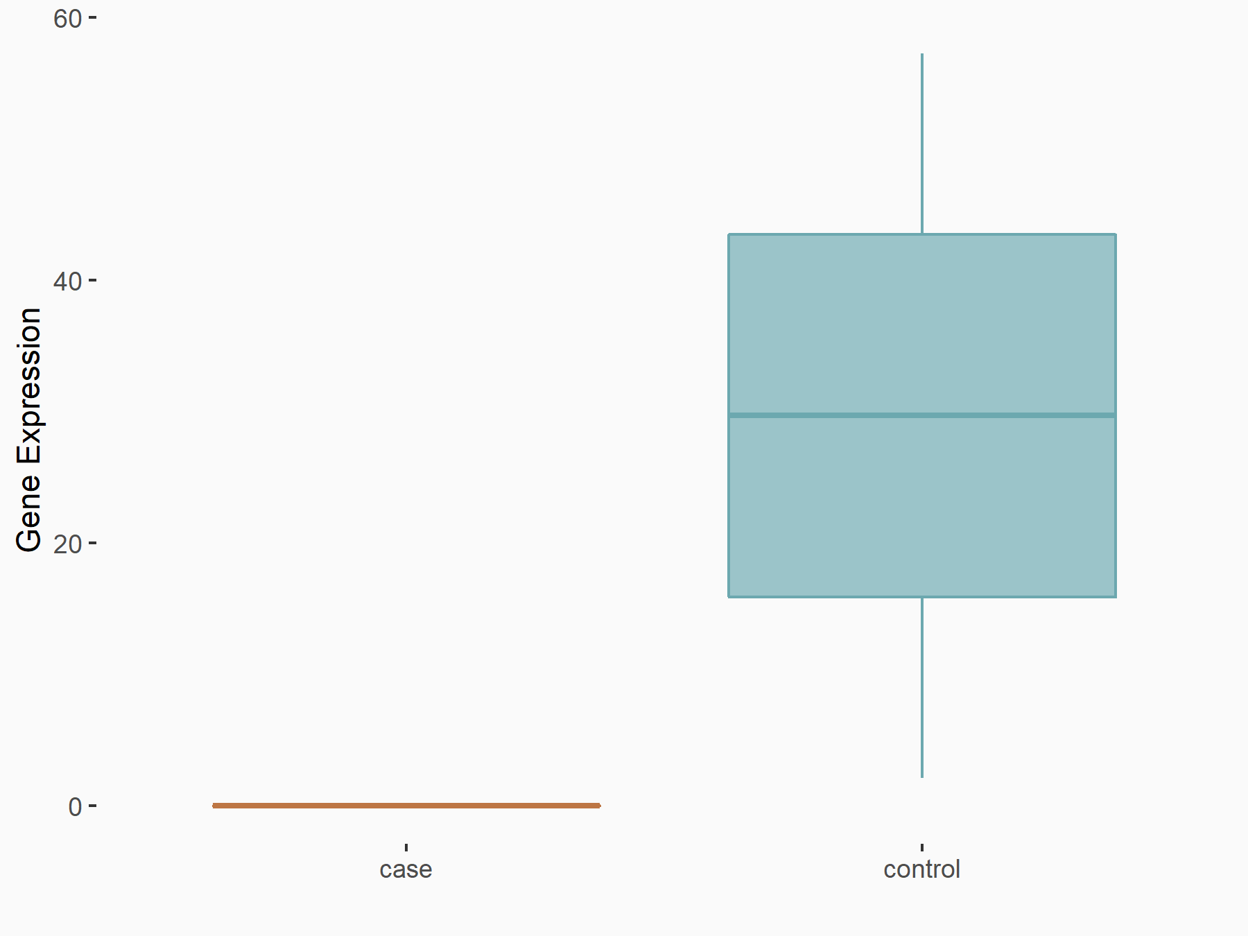
| Representative RNA-seq result indicating the expression of this target gene regulated by FTO | ||
| Cell Line | TPC_1 cell line | Homo sapiens |
|
Treatment: FTO overexpression TPC_1 cells
Control: TPC_1 cells
|
GSE199206 | |
| Regulation |
  |
logFC: -7.40E+00 p-value: 4.06E-02 |
| More Results | Click to View More RNA-seq Results | |
| In total 1 item(s) under this regulator | ||||
| Experiment 1 Reporting the m6A Methylation Regulator of This Target Gene | [1] | |||
| Response Summary | FTO inhibited growth, migration and invasion of GBM cells in vitro and in vivo.decreased FTO expression could induce the downregulation of MTMR3 expression by modulating the processing of pri-miR-10a in an m6A/HNRNPA2B1-dependent manner in GBM cells. Furthermore, the transcriptional activity of FTO was inhibited by the transcription factor Transcription factor PU.1 (SPI1). | |||
| Target Regulation | Down regulation | |||
| Responsed Disease | Glioblastoma | ICD-11: 2A00.00 | ||
| In-vitro Model | U-87MG ATCC | Glioblastoma | Homo sapiens | CVCL_0022 |
| U251 (Fibroblasts or fibroblast like cells) | ||||
| U-118MG | Astrocytoma | Homo sapiens | CVCL_0633 | |
| LN-229 | Glioblastoma | Homo sapiens | CVCL_0393 | |
| A-172 | Glioblastoma | Homo sapiens | CVCL_0131 | |
| In-vivo Model | BALB/c male nude mice were 4 weeks old. GBM cells with stable overexpression or knockdown of FTO and ovNC or shNC were transduced with lentivirus expressing luciferase. The cells were intracranially injected at a density of 5 × 10⁵/10 uL into every mouse to form an orthotopic xenograft model. Coordinates of injection were 1 mm anterior and 2.5 mm right to the bregma, at a depth of 3.5 mm (the right frontal lobes of the mouse). Every 6 days, bioluminescence imaging (IVIS Lumina Series III; PerkinElmer, Waltham, MA) was used to image the mouse. At 8 days, we randomly chose 5 mice from each group to euthanize them, and their brain tissues were fixed with paraformaldehyde for further study. Another 5 mice were used for survival time analysis. For DB2313 (563801; MedKoo) anti-tumor research, male nude mice were subcutaneously injected with 5 × 10⁶ U87MG cells suspended in 0.1 mL PBS. After 7 days, mice were intraperitoneally injected with DB2313 at density of 10 mg/kg/day dissolved in PBS solvent containing 10% DMSO for 7 days. The other group treated with vehicle only was set as the control group. | |||
Brain cancer [ICD-11: 2A00]
| In total 1 item(s) under this disease | ||||
| Experiment 1 Reporting the m6A-centered Disease Response | [1] | |||
| Response Summary | FTO inhibited growth, migration and invasion of GBM cells in vitro and in vivo.decreased FTO expression could induce the downregulation of MTMR3 expression by modulating the processing of pri-miR-10a in an m6A/HNRNPA2B1-dependent manner in GBM cells. Furthermore, the transcriptional activity of FTO was inhibited by the transcription factor Transcription factor PU.1 (SPI1). | |||
| Responsed Disease | Glioblastoma [ICD-11: 2A00.00] | |||
| Target Regulator | Fat mass and obesity-associated protein (FTO) | ERASER | ||
| Target Regulation | Down regulation | |||
| In-vitro Model | U-87MG ATCC | Glioblastoma | Homo sapiens | CVCL_0022 |
| U251 (Fibroblasts or fibroblast like cells) | ||||
| U-118MG | Astrocytoma | Homo sapiens | CVCL_0633 | |
| LN-229 | Glioblastoma | Homo sapiens | CVCL_0393 | |
| A-172 | Glioblastoma | Homo sapiens | CVCL_0131 | |
| In-vivo Model | BALB/c male nude mice were 4 weeks old. GBM cells with stable overexpression or knockdown of FTO and ovNC or shNC were transduced with lentivirus expressing luciferase. The cells were intracranially injected at a density of 5 × 10⁵/10 uL into every mouse to form an orthotopic xenograft model. Coordinates of injection were 1 mm anterior and 2.5 mm right to the bregma, at a depth of 3.5 mm (the right frontal lobes of the mouse). Every 6 days, bioluminescence imaging (IVIS Lumina Series III; PerkinElmer, Waltham, MA) was used to image the mouse. At 8 days, we randomly chose 5 mice from each group to euthanize them, and their brain tissues were fixed with paraformaldehyde for further study. Another 5 mice were used for survival time analysis. For DB2313 (563801; MedKoo) anti-tumor research, male nude mice were subcutaneously injected with 5 × 10⁶ U87MG cells suspended in 0.1 mL PBS. After 7 days, mice were intraperitoneally injected with DB2313 at density of 10 mg/kg/day dissolved in PBS solvent containing 10% DMSO for 7 days. The other group treated with vehicle only was set as the control group. | |||
RNA Modification Sequencing Data Associated with the Target (ID: M6ATAR00408)
| In total 1 m6A sequence/site(s) in this target gene | |||
| mod ID: A2ISITE004717 | Click to Show/Hide the Full List | ||
| mod site | chr11:47360224-47360225:- | [3] | |
| Sequence | TGACCTTGGGCATGCTATGTAGCCGCTCGAAGCCTCAGTGT | ||
| Transcript ID List | ENST00000533030.1; ENST00000378538.7; ENST00000533968.1; rmsk_3543370; ENST00000227163.8 | ||
| External Link | RMBase: RNA-editing_site_21540 | ||
N6-methyladenosine (m6A)
| In total 14 m6A sequence/site(s) in this target gene | |||
| mod ID: M6ASITE005448 | Click to Show/Hide the Full List | ||
| mod site | chr11:47354882-47354883:- | [4] | |
| Sequence | CATTAACCTCCTCCCAAAAAACAAGTAAAGTTATTCTCAAT | ||
| Motif Score | 2.20572619 | ||
| Cell/Tissue List | CD34; MM6 | ||
| Seq Type List | m6A-seq | ||
| Transcript ID List | ENST00000378538.7 | ||
| External Link | RMBase: m6A_site_140015 | ||
| mod ID: M6ASITE005449 | Click to Show/Hide the Full List | ||
| mod site | chr11:47355000-47355001:- | [4] | |
| Sequence | GGACGCCAGCTGGGCGTCAGACCCCACCGGGGCAACCTTGC | ||
| Motif Score | 2.876744048 | ||
| Cell/Tissue List | CD34; LCLs; MM6; CD4T; peripheral-blood; endometrial; NB4 | ||
| Seq Type List | m6A-seq | ||
| Transcript ID List | ENST00000378538.7; ENST00000227163.8 | ||
| External Link | RMBase: m6A_site_140016 | ||
| mod ID: M6ASITE005450 | Click to Show/Hide the Full List | ||
| mod site | chr11:47355037-47355038:- | [4] | |
| Sequence | CCGCTTCGCCTCCCTCCAGGACTCCACCCCGGCTCCCGGAC | ||
| Motif Score | 4.065041667 | ||
| Cell/Tissue List | CD34; GM12878; LCLs; MM6; CD4T; peripheral-blood; endometrial; NB4 | ||
| Seq Type List | m6A-seq | ||
| Transcript ID List | ENST00000378538.7; ENST00000227163.8 | ||
| External Link | RMBase: m6A_site_140017 | ||
| mod ID: M6ASITE005451 | Click to Show/Hide the Full List | ||
| mod site | chr11:47355120-47355121:- | [5] | |
| Sequence | TCCCGGGGCCCAGAGGCAGGACTGTGGCGGGCCGGGCCTCG | ||
| Motif Score | 4.065041667 | ||
| Cell/Tissue List | HEK293T; GM12878; LCLs; MM6; CD4T; peripheral-blood; endometrial; NB4 | ||
| Seq Type List | MeRIP-seq; m6A-seq | ||
| Transcript ID List | ENST00000533030.1; ENST00000227163.8; ENST00000378538.7 | ||
| External Link | RMBase: m6A_site_140018 | ||
| mod ID: M6ASITE005452 | Click to Show/Hide the Full List | ||
| mod site | chr11:47355155-47355156:- | [5] | |
| Sequence | TAAGCCCTCGCCCGGCCCGGACACAGGGAGGACGCTCCCGG | ||
| Motif Score | 3.643047619 | ||
| Cell/Tissue List | HEK293T; GM12878; LCLs; MM6; CD4T; peripheral-blood; endometrial; NB4 | ||
| Seq Type List | MeRIP-seq; m6A-seq | ||
| Transcript ID List | ENST00000227163.8; ENST00000378538.7; ENST00000533030.1 | ||
| External Link | RMBase: m6A_site_140019 | ||
| mod ID: M6ASITE005453 | Click to Show/Hide the Full List | ||
| mod site | chr11:47355455-47355456:- | [4] | |
| Sequence | CATCTGGTGGGTGGACAAGGACAAGGGCACCTTCCAGTTCT | ||
| Motif Score | 3.643047619 | ||
| Cell/Tissue List | CD34; MM6; CD4T; peripheral-blood; endometrial; NB4 | ||
| Seq Type List | m6A-seq | ||
| Transcript ID List | ENST00000378538.7; ENST00000227163.8; ENST00000533030.1 | ||
| External Link | RMBase: m6A_site_140020 | ||
| mod ID: M6ASITE005454 | Click to Show/Hide the Full List | ||
| mod site | chr11:47355461-47355462:- | [4] | |
| Sequence | GGACAGCATCTGGTGGGTGGACAAGGACAAGGGCACCTTCC | ||
| Motif Score | 3.643047619 | ||
| Cell/Tissue List | CD34; MM6; CD4T; peripheral-blood; endometrial; NB4 | ||
| Seq Type List | m6A-seq | ||
| Transcript ID List | ENST00000533030.1; ENST00000378538.7; ENST00000227163.8 | ||
| External Link | RMBase: m6A_site_140021 | ||
| mod ID: M6ASITE005455 | Click to Show/Hide the Full List | ||
| mod site | chr11:47355479-47355480:- | [4] | |
| Sequence | CCGCAGCGGCGACATGAAGGACAGCATCTGGTGGGTGGACA | ||
| Motif Score | 3.643047619 | ||
| Cell/Tissue List | CD34; MM6; CD4T; peripheral-blood; endometrial; NB4 | ||
| Seq Type List | m6A-seq | ||
| Transcript ID List | ENST00000533030.1; ENST00000378538.7; ENST00000227163.8 | ||
| External Link | RMBase: m6A_site_140022 | ||
| mod ID: M6ASITE005456 | Click to Show/Hide the Full List | ||
| mod site | chr11:47355506-47355507:- | [4] | |
| Sequence | CCTGTACCAGTTCCTGTTGGACCTGCTCCGCAGCGGCGACA | ||
| Motif Score | 3.622404762 | ||
| Cell/Tissue List | CD34; MM6; peripheral-blood; endometrial; NB4 | ||
| Seq Type List | m6A-seq | ||
| Transcript ID List | ENST00000378538.7; ENST00000533030.1; ENST00000227163.8 | ||
| External Link | RMBase: m6A_site_140023 | ||
| mod ID: M6ASITE005457 | Click to Show/Hide the Full List | ||
| mod site | chr11:47358846-47358847:- | [4] | |
| Sequence | CTGGGCTCCTGCCTGGGGAGACAGGTGGGGCACCTGGCCCT | ||
| Motif Score | 2.897386905 | ||
| Cell/Tissue List | CD34; MM6; peripheral-blood; endometrial | ||
| Seq Type List | m6A-seq | ||
| Transcript ID List | ENST00000227163.8; ENST00000533030.1; ENST00000378538.7; ENST00000533968.1 | ||
| External Link | RMBase: m6A_site_140024 | ||
| mod ID: M6ASITE005458 | Click to Show/Hide the Full List | ||
| mod site | chr11:47359979-47359980:- | [4] | |
| Sequence | GTTCGAGAGCTTCGCCGAGAACAACTTCACGGAGCTCCAGA | ||
| Motif Score | 2.951386905 | ||
| Cell/Tissue List | CD34; peripheral-blood; endometrial | ||
| Seq Type List | m6A-seq | ||
| Transcript ID List | ENST00000533968.1; ENST00000378538.7; ENST00000227163.8; ENST00000533030.1 | ||
| External Link | RMBase: m6A_site_140025 | ||
| mod ID: M6ASITE005459 | Click to Show/Hide the Full List | ||
| mod site | chr11:47360027-47360028:- | [4] | |
| Sequence | CCCCACAGACCATTACTGGGACTTCCACCCCCACCACGTGC | ||
| Motif Score | 4.065041667 | ||
| Cell/Tissue List | CD34; peripheral-blood | ||
| Seq Type List | m6A-seq | ||
| Transcript ID List | ENST00000227163.8; ENST00000533968.1; ENST00000378538.7; ENST00000533030.1 | ||
| External Link | RMBase: m6A_site_140026 | ||
| mod ID: M6ASITE005460 | Click to Show/Hide the Full List | ||
| mod site | chr11:47375718-47375719:- | [4] | |
| Sequence | CTCTCAGCAGCCATCAGAAGACCTGGTGCCCTATGACACGG | ||
| Motif Score | 2.876744048 | ||
| Cell/Tissue List | CD34; MM6; CD4T; peripheral-blood; NB4 | ||
| Seq Type List | m6A-seq | ||
| Transcript ID List | ENST00000378538.7; ENST00000533968.1; ENST00000533030.1; ENST00000227163.8 | ||
| External Link | RMBase: m6A_site_140027 | ||
| mod ID: M6ASITE005461 | Click to Show/Hide the Full List | ||
| mod site | chr11:47378536-47378537:- | [6] | |
| Sequence | GCCCTTCGATAAAATCAGGAACTTGTGCTGGCCCTGCAATG | ||
| Motif Score | 3.373380952 | ||
| Cell/Tissue List | MM6; CD4T | ||
| Seq Type List | m6A-seq | ||
| Transcript ID List | ENST00000378538.7 | ||
| External Link | RMBase: m6A_site_140028 | ||
References